
Fascia is a specialized connective tissue wrapping muscles, bones, and organs, classified into four main types based on location and structure: superficial, deep, visceral, and parietal. These layers allow for structural integrity, sliding, and gliding, crucial for movement, and provide support for blood vessels and nerves throughout the body.
The Four Primary Types of Fascia
Superficial fascia. Located directly under the skin in the hypodermis, this layer contains fat and elastic fibers. It acts as a covering for the body, allowing skin to move over deeper tissues.
Deep fascia. A dense, fibrous connective tissue that surrounds muscles, bones, nerves, and blood vessels. It acts like a stocking, separating muscles into compartments and providing a base for muscle attachment. Key examples include the fascia lata in the thigh and thoracolumbar fascia in the back.
Visceral fascia. Also known as subserous fascia, it covers organs (viscera) and suspends them within their cavities (e.g., in the chest or abdomen).
Parietal fascia. Lines the walls of body cavities (such as the pelvis or abdomen), lying outside the parietal layer of the serous membranes.
Intermediate Layers
Loose Fascia/Fascial Interface: An intermediate layer that often exists between superficial and deep fascia, facilitating a “sliding and gliding” motion between layers.
Key Functions
Movement and stability. Provides structure, reduces friction, and enables smooth movement between muscles.
Protection. Wraps muscles and organs, shielding them from trauma.
Force transmission. Allows for effective force distribution during movement.
_____________________________
What is Fascia?
Fascia is a tough connective tissue that spreads throughout the entire body in a three-dimensional pattern from head to foot without interruption. Like a spider web, the fascia covers the muscles, bones, nerves, organs, arteries, and veins down to the cellular level. Each part of the entire body is connected to every other part by the fascia, much like the yarn in a sweater. When one area of the sweater is pulled, it affects another area due to the thread connection. This can also be true with fascia; when one area of the body feels pain, the base of the pain may be coming from another region within the body. Therefore, it’s important that you address not only the area of pain, for example, the shoulder but what may be the cause of the pain – abdominal scars, poor posture, etc.
Trauma, surgery, inflammation, poor posture, scars, bracing against pain, and even anxiety can bind down the fascia and solidify, causing abnormal pressure of approximately 2,000 pounds per square inch on any or all of these body parts. This can result in pain, lack of motion, and dehydration of the fluid component of the fascia causing it to lose its relaxed and wavy configuration and become like a straight-jacket.
Since many current standard tests such as x-rays, myelograms, CAT scans and EMG’s do not show fascial restrictions, many people suffering with pain may have fascial restrictions that need to be addressed. Currently, elastography, the study of addressing the elasticity of tissue, may help further address localization of fascial restrictions and help to better diagnose individuals experiencing pain.
Fascial restrictions can be cumulative. This means that some of the trauma could have been small and accumulated over time – even a fall when you were 2 years old can result in a pain response at a later time.
What is Myofascial Release?
Myofascial release therapy is a safe effective, hands-on technique. Myofascial release techniques involve a gentle, sustained, low load pressure into the restricted areas for sometimes up to as much as 5 minutes or more to one area. Myofascial therapy performed by a licensed therapist is used not to just stretch the tissue but elongate it (Fig. 1-4). Fascia tends to resist suddenly applied force and does not generally respond well to quick stretches, therefore, myofascial release therapy can be very beneficial in treating myofascial pain syndrome.
Myofascial therapy is applied by a licensed therapist
Figure 1: Myofascial therapy is applied by a licensed therapist
Myofascial therapy is applied by a licensed therapist
Figure 2: Myofascial therapy is applied by a licensed therapist
Myofascial therapy is applied by a licensed therapist
Figure 3: Myofascial therapy is applied by a licensed therapist
Myofascial therapy is applied by a licensed therapist
Figure 4: Myofascial therapy is applied by a licensed therapist
Each person with myofascial pain is looked at as a totally unique individual. Their treatment is focused specifically on their issues, not only addressing the area of pain but assessing and addressing the cause of the pain. It is not a “no pain no gain” type of treatment. Myofascial release therapy, a type of neuromuscular therapy, allows you to experience “therapeutic pain” which allows old tissue memory of injury or trauma to release. Many of us relate pain to injury but, in this case, when treatment and self-myofascial release exercises are done and you feel a type of pain or softening, it is probably your body’s way of telling you that what you are doing is appropriate.
Self-myofascial release is the treatment of incorporating various techniques which may include the use of a tacky ball and foam roller into your exercise program (Fig. 5-7). The fascial release effects provided by the use of a foam roller (typically referred to as foam roller therapy) in conjunction with your own body weight can facilitate release in multiple areas of your body. By rolling slowly over the foam roller and the targeted area, the foam roller stretches and elongates the fascia and actually allows you to reach deep muscle tissues.
Tacky ball and foam roller exercises
Figure 5: Tacky ball exercise
Tacky ball and foam roller exercises
Figure 6: Foam roller exercise
Tacky ball and foam roller exercises
Figure 7: Foam roller exercise
With myofascial release or any type of alternative medicine therapy, it’s not always enough to simply go through the motions of the exercises but to fully participate by bringing ones full attention into the body or location of pain. Using one’s breath is also important to help soften or melt away the area of tightness or pain.
What can you expect with Myofascial Release?
When fascia releases, it can feel like taffy softening. Sometimes people may feel cold or heat, tingling or buzzing, burning, or a sense of water or air moving through the area. They may also notice a sensation in an area other than that being treated. This is called the “fascial voice” and is your body’s way of telling you that this area also needs to be elongated and given attention. People will generally notice an increase in their motion and increased function with less pain.
It is always important to drink plenty of water after treatment to help flush out metabolic wastes and re-hydrate the tissue.
Many of our therapists at the Hand to Shoulder Center of Wisconsin have been trained in variable myofascial release techniques which they incorporate in their treatment with simple hand, wrist, elbow, and shoulder injuries to complex traumas and pain of unknown cause.
Myofascial release is done in conjunction with patient participation through a home therapy program, understanding of their pain, and activity and work station modifications to reduce their physical and mental stressors. Our goal is to provide the patient with the tools to self-manage their pain and resume activities that they could no longer participant in and enjoy!
_________
Bordoni B, Mahabadi N, Varacallo MA. Anatomy, Fascia. [Updated 2023 Jul 17]. In: StatPearls [Internet]. Treasure Island (FL): StatPearls Publishing; 2025 Jan-. Available from: https://www.ncbi.nlm.nih.gov/books/NBK493232/
Bordoni B, Zanier E. Skin, fascias, and scars: symptoms and systemic connections. J Multidiscip Healthc. 2013 Dec 28;7:11-24. doi: 10.2147/JMDH.S52870. PMID: 24403836; PMCID: PMC3883554.
Fibrosis and densification: Anatomical vs functional alteration of the fascia
Pavan, Piero et al.
Journal of Bodywork and Movement Therapies, Volume 20, Issue 1, 151
https://www.bodyworkmovementtherapies.com/article/S1360-8592(15)00199-0/abstract
Kodama, Y., Masuda, S., Ohmori, T., Kanamaru, A., Tanaka, M., Sakaguchi, T., & Nakagawa, M. (2023). Response to Mechanical Properties and Physiological Challenges of Fascia: Diagnosis and Rehabilitative Therapeutic Intervention for Myofascial System Disorders. Bioengineering (Basel, Switzerland), 10(4), 474. https://doi.org/10.3390/bioengineering10040474
Lv, Y., & Yin, Y. (2024). A Review of the Application of Myofascial Release Therapy in the Treatment of Diseases. Journal of multidisciplinary healthcare, 17, 4507–4517. https://doi.org/10.2147/JMDH.S481706
https://dx.doi.org/10.16965/ijar.2020.237
Site-specific fascia tuning pegs and places of perilous passage myofascial considerations in upper extremity entrapment neuropathies: A clinical anatomists view
John Sharkey MSc.
10.16965/ijar.2020.237
https://www.youtube.com/watch?v=_FtSP-tkSug
https://www.gilhedley.com/0-ce-whats-the-fuzz-course-description
Bordoni B, Mahabadi N, Varacallo MA. Anatomy, Fascia. [Updated 2023 Jul 17]. In: StatPearls [Internet]. Treasure Island (FL): StatPearls Publishing; 2025 Jan-. Available from: https://www.ncbi.nlm.nih.gov/books/NBK493232/
Stecco C, Fede C, Macchi V, Porzionato A, Petrelli L, Biz C, Stern R, De Caro R. The fasciacytes: A new cell devoted to fascial gliding regulation.Clin Anat. 2018 Jul;31(5):667-676.
Stecco C, Macchi V, Barbieri A, Tiengo C, Porzionato A, De Caro R. Hand fasciae innervation: The palmar aponeurosis. Clin Anat. 2018 Jul;31(5):677-683.
Stecco C, Azzena GP, Macchi V, Porzionato A, Behr A, Rambaldo A, Tiengo C, De Caro R. Rectus abdominis muscle innervation: an anatomical study with surgical implications in diep flap harvesting. Surg Radiol Anat. 2017 Nov 10.
Stecco C, Fantoni I, Macchi V, Del Borrello M, Porzionato A, Biz C, De Caro R. The role of fasciae in Civinini-Morton’s syndrome. J Anat. 2015 Nov;227(5):654-64.
Stecco C, Sfriso MM, Porzionato A, Rambaldo A, Albertin G, Macchi V, De Caro R. Microscopic anatomy of the visceral fasciae.J Anat. 2017 Jul;231(1):121-128.
Stecco C, Cappellari A, Macchi V, Porzionato A, Morra A, Berizzi A, De Caro R. The paratendineous tissues: an anatomical study of their role in the pathogenesis of tendinopathy. Surg Radiol Anat. 2014 Aug; 36(6):561-72.
Stecco A, Stern R, Fantoni I, De Caro R, Stecco C. Fascial Disorders: Implications for Treatment. PM R. 2015 Jun 14. pii: S1934-1482(15)00292-0.
P.G. Pavan, A. Stecco, R. Stern, C. Stecco, Painful connections: densification versus fibrosis of fascia, Current Pain and Headache Reports, 2014 Aug, 18(8):441, doi: 10.1007/s11916-014-0441-4, PubMed PMID: 25063495.
T. Luomala, M. Pihlman, J. Heiskanen, C. Stecco, Case study: could ultrasound and elastography visualized densified areas inside the deep fascia?, Journal of Bodywork and Movement Therapies, 2014 Jul, 18(3):462-8, doi: 10.1016/j.jbmt.2013.11.020, PubMed PMID: 25042323.
C. Stecco, A. Cappellari, V. Macchi, A. Porzionato, A. Morra, A. Berizzi, R. De Caro, The paratendineous tissues: an anatomical study of their role in the pathogenesis of tendinopathy, Surgical and Radiologic Anatomy, 2014 Aug, 36(6):561-72, doi: 10.1007/s00276-013-1244-8, PubMed PMID: 24318515.
Yes, fascia has significantly more nerve endings than most other tissues in the body, including muscle, making it considered the body’s richest sensory organ; meaning it contains a higher density of nerve endings compared to other tissues, allowing it to provide extensive sensory information about movement and position.
Key points about fascia and nerve endings:
High nerve density:
Fascia can have 6 to 10 times more nerve endings than muscle tissue.
Proprioception:
These nerve endings are crucial for proprioception, which is the body’s awareness of its position in space.
Pain perception:
Fascia also contains nociceptors, which are responsible for detecting pain signals.
Sensory organ:
Due to its extensive nerve supply, fascia is often referred to as the body’s largest sensory organ.
Therefore, the researchers believe that the nervous system resides in Fascia
Therefore, the researchers believe that the nervous system resides in Fascia
Recent research shows that the Fascial network contains around 250 million free nerve endings and pain receptors (nociceptors), compared to the skin’s 200 million and the eye’s 126 million visual receptors. (If you also count the skin as Fascia, it would be 450 million free nerve endings). This means that Fascia is our richest innervated organ, our largest sensory organ! Fascia communicates with all other organs and cells in the body. It sends signals and receives information in several different ways, to and from other organs and tissues in the body.
Is it only through the nervous system and hormones that the body communicates? No, new knowledge shows that in addition to the nervous system’s signals, there is communication that goes much faster than the fastest nerve cell! Fascia consists largely of a fluid ground substance, interstitium, which contains a multitude of large water-holding molecules, such as hyaluronic acid and other molecules, between networks of collagen protein fibers. In this flow, a multitude of signaling molecules circulate in Fascia, with a multitude of information. The slightest movement is conveyed to the body’s cells in the collagen fiber network, and weak currents are formed by the movement.
Int J Mol Sci. 2022 May 18;23(10):5674. doi: 10.3390/ijms23105674
Fascial Innervation: A Systematic Review of the Literature
Vidina Suarez-Rodriguez 1,*, Caterina Fede 2, Carmelo Pirri 2, Lucia Petrelli 2, Juan Francisco Loro-Ferrer 3, David Rodriguez-Ruiz 4, Raffaele De Caro 2, Carla Stecco 2
https://pmc.ncbi.nlm.nih.gov/articles/PMC9143136/
. Since then, several investigations have been performed and published about fascial innervation, proposing that the fasciae may be considered our largest sensory organ given its complete surface area, as well as participating actively in proprioception and nociception.
https://www.muscleiq.com/post/is-fascia-another-communication-system-inside-our-body
Is Fascia Another Communication System Inside our body?
Fascia is more than a supportive springy wrap, though. It also contains blood vessels and sensory receptors. The fascia has between 6 to 10 times more nerve endings than muscle.
Loosening fascia can help increase range of motion before exercise and can limit soreness after exercise.
https://www.healthline.com/health/dermatome
The 30 Dermatomes Explained and Located
https://www.myofascialmississauga.com/blog/fascia-and-nerve-dysfunction-dams-in-the-river
Pratt R. L. (2021). Hyaluronan and the Fascial Frontier. International journal of molecular sciences, 22(13), 6845. https://doi.org/10.3390/ijms22136845
_____
Lipedema is an adipofascial disorder that can cause chronic pain and swelling. It can involve abnormalities in the superficial fascia, which can lead to pain and inflammation.
How does lipedema affect fascia?
Clogged fascia: Poor lymph drainage can cause fibrinogen to build up in the lymphatic vessels, clogging the fascia.
Broken fascia lines: Visible breaks in the fascia lines can be seen on ultrasounds.
Fibrosis: The inflammatory process of lipedema can create nodules and hard fibrosis around the subcutaneous adipose tissue cells.
How can fascia training help with lipedema?
Fascia training can help loosen clogged-up fascia tissue and soften the fascia.
Fascia massage rollers can help loosen the tissue.
Other treatments for lipedema:
Complete decongestive therapy (CDT) can help decongest swelling, encourage lymphatic vessel pumping, and decrease pain.
Compression garments can help with cosmetic reasons.
Bariatric surgery can help with overall glucometabolic aspects.
Liposuction can improve pain, muscle cramps, tightening, itching, edema, bruising, and cosmetic appearance.
Lipedema mainly affects women and may be related to female hormonal changes.
Biomedicines. 2022 Nov 30;10(12):3081. doi: 10.3390/biomedicines10123081
Lipedema: Insights into Morphology, Pathophysiology, and Challenges
Ankita Poojari 1,†, Kapil Dev 1,†, Atefeh Rabiee 1,*
https://pmc.ncbi.nlm.nih.gov/articles/PMC9775665/
https://www.lipedema.com/lipedema-is-not-just-fat
Superficial fascia abnormalities, visible as breaks in the fascia lines on ultrasound, appear to be common in lipedema and may be improved following therapy. See for example Figure 2 in [56]. Abnormalities of the superficial fascia can contribute to chronic fatigue, fibromyalgia, pain, and inflammation.[8]
Long-distance Interstitial fluid flows through fibrous matrices within LCT and superficial fascia which are relatively independent of the vascular circulation have been identified and mapped.
Lipedema and exercise
Is lipedema resistant to exercise?
https://www.ofa-bamberg.com/en/knowledge/therapy/cdp/lipedema-and-sport/
Fascia training: Fasciae are part of the connective tissue and surround joints, muscles and bones as well as the lymphatic vessels like a shell. They consist of water, collagen and adhesive matter. If the lymph drainage does not function properly (which often occurs in the case of lipedema), fibrinogen will accumulate in the lymphatic vessels, which can cause the fasciae to clog. Clogged-up fascia tissue can be a cause of the lipedema pain. Fascia training is designed to loosen this clogged-up connective tissue again and to soften the fasciae. Fascia massage rollers in different sizes and with varying intensities can help to loosen the tissue. Please note: Using these rollers can be quite painful initially, until the clogged-up fasciae are starting to loosen up.
George T, De Jesus O. Physiology, Fascia. [Updated 2023 Mar 12]. In: StatPearls [Internet]. Treasure Island (FL): StatPearls Publishing; 2026 Jan-. Available from: https://www.ncbi.nlm.nih.gov/books/NBK568725/
Fascial nomenclature: Update on related consensus process
Robert Schleip, Gil Hedley, Can A. Yucesoy
First published: 10 June 2019 https://doi.org/10.1002/ca.23423
Clinical Anatomy.
Donahue, P., Crescenzi, R., Petersen, K., Garza, M., et al. (2022). Physical therapy in women with early stage lipedema: Potential impact of multimodal manual therapy, compression, exercise, and education interventions. Lymphatic Research and Biology.
Gordon, C. M., Lindner, S. M., Birbaumer, N., Montoya, P., Ankney, R. L., & Andrasik, F. (2018). Self-Myofascial Vibro-Shearing: a Randomized Controlled Trial of Biomechanical and Related Changes in Male Breakdancers. Sports medicine – open, 4(1), 13. https://doi.org/10.1186/s40798-018-0128-1
Stecco, A., Giordani, F., Fede, C., Pirri, C., De Caro, R., & Stecco, C. (2023). From Muscle to the Myofascial Unit: Current Evidence and Future Perspectives. International journal of molecular sciences, 24(5), 4527. https://doi.org/10.3390/ijms24054527
George T, De Jesus O. Physiology, Fascia. [Updated 2023 Mar 12]. In: StatPearls [Internet]. Treasure Island (FL): StatPearls Publishing; 2025 Jan-. Available from: https://www.ncbi.nlm.nih.gov/books/NBK568725/
Fede, C., Clair, C., Pirri, C., Petrelli, L., Zhao, X., Sun, Y., Macchi, V., & Stecco, C. (2025). The Human Superficial Fascia: A Narrative Review. International journal of molecular sciences, 26(3), 1289. https://doi.org/10.3390/ijms26031289
Yildizgoren, M., & Dede, B., (2024). The role of fascia in myofascial pain syndrome: A look at Cinderella tissue. Cam & Sakura Medical Journal.
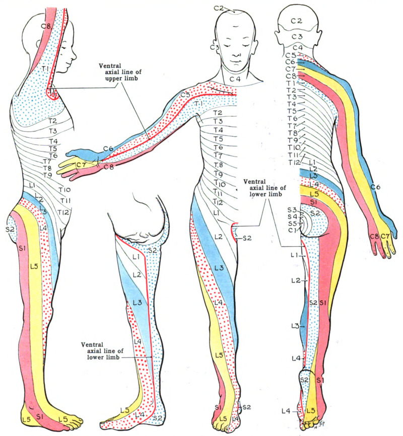
Whitman, P.A., et al. (2023). Anatomy, Skin, Dermatomes
Whitman PA, Launico MV, Adigun OO. Anatomy, Skin, Dermatomes. [Updated 2023 Oct 24]. In: StatPearls [Internet]. Treasure Island (FL): StatPearls Publishing; 2025 Jan-. Available from: https://www.ncbi.nlm.nih.gov/books/NBK535401/
FASCIA AND CELLULITE
Adamidis, N., Papalexis, P., & Adamidis, S. (2024). Exploring the Link Between Metabolic Syndrome and Cellulite. Cureus, 16(6), e63464. https://doi.org/10.7759/cureus.63464
Shafaroodi, H., Barati, S., Ghasemi, M., Almasirad, A., & Moezi, L. (2016). A role for ATP-sensitive potassium channels in the anticonvulsant effects of triamterene in mice. Epilepsy research, 121, 8–13. https://doi.org/10.1016/j.eplepsyres.2016.01.003
Singh, A., Gupta, A., Chowdhary, M. and Brahmbhatt, H.D. (2021), Integrated analysis of miRNA-mRNA networks reveals a strong anti-skin cancer signature in vitiligo epidermis. Exp Dermatol, 30: 1309-1319. https://doi.org/10.1111/exd.14317
https://ibspot.com/us/products/beurer-cm100-cellulite-releazer-helps-reduce-the-appearance-of-cellulite-cellulite-massager-waterproof-massager-roller-cellulite-blast-treatment-white-teal
Additional Information
Fascia Lines are the intersections of the body
https://my.clevelandclinic.org/health/body/23251-fascia
Fascia as a flow – a new way of explaining the body’s function
Fascia as a flow – a new way of explaining the body’s function
Fascia is the largest organ in the body
https://www.ncbi.nlm.nih.gov/books/NBK493232/
https://www.sciencedirect.com/science/article/pii/S235230931930015X